
Eyelid ptosis: when a drooping eyelid is not just tiredness
An eyelid covering part of the pupil is not corrected by removing skin. What causes ptosis, why it is urgent in children, and how the surgical technique is chosen.
By Dr. Miguel Ángel Olivardía Moreno · · Updated 3 de agosto de 2026
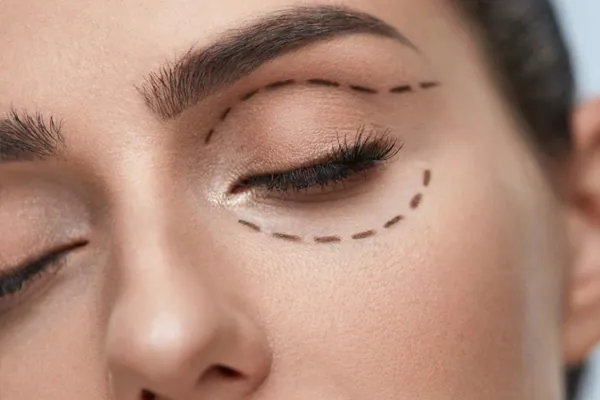
Eyelid ptosis is the drooping of the upper eyelid below its normal position. It is not the same as excess skin, even though from the outside they can look alike: in ptosis the eyelid margin itself is low; in dermatochalasis what is in excess is skin hanging over a margin that is where it should be.
Telling one from the other is the first step, because each is corrected with a different operation.
How it is recognised
The objective sign is the position of the eyelid margin relative to the pupil. But there are indirect signs that patients do notice:
- One eye looks smaller or more «sleepy» than the other.
- Constantly raising the eyebrows to clear the vision, which over the years etches forehead lines.
- Tilting the head back in order to look straight ahead.
- Visual fatigue at the end of the day.
- Checking old photographs and finding the asymmetry was not there before.
The causes
Aponeurotic (the most frequent in adults). With age, the aponeurosis of the levator muscle becomes detached or thins. The muscle works well, but its anchorage to the eyelid has come loose. It is the typical cause of ptosis in older adults.
Congenital. The levator muscle did not develop properly. It is present from birth and its management follows a different logic, explained below.
Mechanical. An eyelid tumour, inflammation or the weight of excess tissue drags the eyelid downwards.
Neurogenic. A disturbance in the nerve supply to the muscle, as in third cranial nerve palsy or Horner's syndrome.
Myogenic. Diseases affecting the muscle or the neuromuscular junction, such as myasthenia gravis, in which the ptosis characteristically fluctuates and worsens towards the end of the day.
Post-surgical or traumatic. After previous eye surgery or an injury.
Identifying the cause is not an academic exercise: it determines whether treatment is surgical, whether neurological investigation is needed first, or whether the problem is systemic.
In children, the clock is running
Congenital ptosis deserves a paragraph of its own. During the first years of life the visual system is being built, and that process depends on both eyes sending sharp images to the brain.
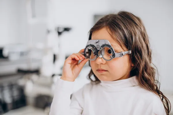
If the eyelid covers the visual axis, the brain begins to ignore the information from that eye. The result is amblyopia: a loss of vision that is not in the eye but in the processing, and which past a certain age no longer fully recovers, even if the ptosis is corrected.
That is why significant congenital ptosis is assessed early. The decision about when to operate depends on the degree of obstruction, on levator muscle function, and on whether the child is already developing amblyopia or a compensatory head tilt.
What is measured before surgery
The surgical technique is not chosen by preference but by measurement. The essentials:
- Levator function: how much travel the eyelid has between extreme downgaze and upgaze.
- Degree of ptosis: how many millimetres the margin sits below normal.
- Bell's phenomenon: whether the eye rotates upwards on closing. This is what protects the cornea if closure is incomplete after surgery.
- State of the ocular surface: significant dry eye limits how far the eyelid can safely be raised.
- Ruling out neurological or myogenic causes before scheduling any surgery.
The techniques
When levator function is good, the aponeurosis is addressed: it is reattached or shortened. This is the most common situation in adult ptosis and the one that usually gives the most predictable results.
When levator function is poor or absent, there is no useful muscle to reinforce. There, frontalis suspension is used: the eyelid is connected to the forehead with a suspension material, so that the patient lifts the eyelid using the brow. It is the usual technique in severe congenital ptosis.
A delicate balance
Correcting ptosis means finding a middle point between two opposite risks. Lifting too little leaves the problem unsolved. Lifting too much stops the eyelid closing properly, and an eye that does not close fully exposes the cornea, dries out and can ulcerate.
That balance is the reason the preoperative assessment is so meticulous, and why a proportion of cases — especially those with very reduced muscle function — may need a later adjustment. It is better to know that from the outset.