




Squamous cell carcinoma of the conjunctiva
An uncommon, slow-growing tumour, more frequent in tropical countries because of ultraviolet radiation. What it is, how it is diagnosed and why follow-up matters as much as the surgery.
By Dr. Miguel Ángel Olivardía Moreno · · Updated 3 de agosto de 2026
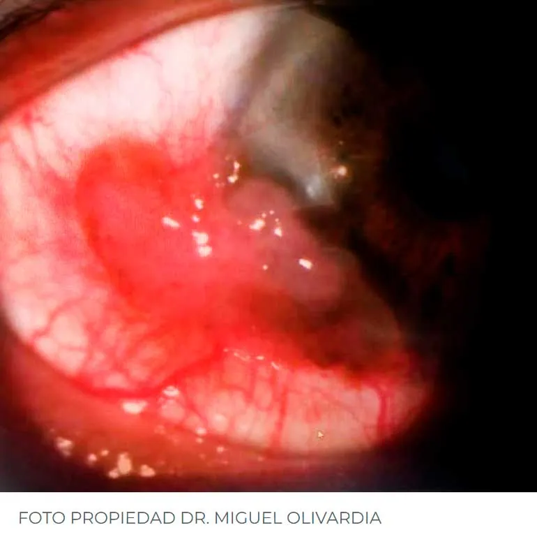
Squamous cell carcinoma of the conjunctiva is a rare, slow-growing, low-grade malignant tumour that can arise de novo or from a pre-existing conjunctival or corneal intraepithelial neoplasia. It occurs more frequently in patients with xeroderma pigmentosum and with AIDS.
The incidence varies between 0.13 and 1.9 cases per 100,000 population and is more frequent in tropical countries because of ultraviolet B radiation at 290–320 nm, where macroinvasive forms and metastases are also frequent, the latter determined by delay in diagnosis or treatment.
How it presents
Clinically, squamous cell carcinoma is painless. It presents between the sixth and seventh decade of life and appears as an exophytic, sessile or pedunculated lesion in the interpalpebral exposure zone, of variable appearance and frequently near the limbus. It may arise on pre-existing lesions such as carcinoma in situ, solar keratosis or epithelial dysplasia.
Unlike the lesions grouped under the generic name of conjunctival intraepithelial neoplasia or conjunctival dysplasia, squamous carcinoma shows, by definition, breach of the basement membrane and infiltration of the corion, which determines a change in therapeutic approach.
Dysplasia is classified by the degree of epithelial involvement: mild, or conjunctival intraepithelial neoplasia (CIN) grade I, confined to the lower third of the epithelial thickness; moderate dysplasia (CIN II), extending to the middle third; and severe dysplasia (CIN III), reaching the upper third.
Diagnosis
Diagnosis is made by biopsy with wide excision. Where ocular invasion is suspected it can be assessed by ultrasound, and for orbital assessment with imaging, preferably contrast MRI.
The differential diagnosis includes chemical burns, dermoid, limbal melanoma and pterygium.
Pattern of invasion
Intraocular invasion follows a typical pattern: tumour cells progress through the scleral channels of the limbus and the ciliary arteries to the trabecular meshwork and, from there, to the root of the iris, the ciliary body and the subretinal space.
There may be invasion of the aqueous humour by tumour cells, diagnosable by anterior chamber tap and cytological study with Papanicolaou staining. The retina, vitreous and lens are not usually affected.
Orbital invasion occurs by extension, and distant metastases appear most frequently in the preauricular, submandibular and anterior cervical nodes, and in the parotid gland. Cases of bone and lung metastases have been described.
Two less common variants
Spindle cell squamous carcinoma. It is rare and much more aggressive. It presents as flat lesions with marked aggressiveness and a tendency to intraocular penetration, sometimes mimicking peripheral corneal ulcers or pterygium. Histologically the cells are spindle-shaped and pleomorphic, with a hyperchromatic nucleus, at times hard to distinguish from fibroblasts, and can be wrongly diagnosed as fibrous histiocytomas or fibrosarcomas. Immunohistochemical studies positive for cytokeratins confirm the epithelial nature of the tumour.
Mucoepidermoid carcinoma. Also rare, it usually appears in elderly patients. It is more aggressive than the spindle cell variant and tends to invade the globe and the orbit. It usually appears in the conjunctival fornix, with a yellowish globular component due to the mucus-secreting cells.
Treatment
Conjunctival lesions can be treated by histologically controlled resection, with or without sclerectomy, additionally using cryotherapy as adjuvant management. In some cases sclerotomy or superficial lamellar keratectomy may be needed.
The recurrence rate after excision is 30 %, and falls below 5 % if a clear histological margin is obtained. In some cases local excision combined with brachytherapy is necessary. Enucleation is reserved for cases of intraocular invasion; where there is orbital invasion, exenteration may be needed, and in extensive lesions radical radiotherapy must be considered.
Topical chemotherapy with mitomycin-C or 5-fluorouracil may be indicated for relapses or as primary treatment in selected early cases and after excisional biopsy. If the patient cannot tolerate extensive surgery, palliative treatment is considered.
Follow-up is not optional
Follow-up after treatment must be close, in order to spot any sign of recurrence and treat it aggressively if it appears.
Occasionally a pyogenic granuloma may appear after excision in areas where the sclera has no covering. It responds rapidly to treatment with topical steroid and must be distinguished from a recurrent tumour.
The main complication is recurrence, typically during the first year after excision, although it can present some time later. Intraocular invasion has been reported in 2 to 8 % of cases and orbital invasion in 12 to 16 %.
The prognosis is reasonably good where excision of the lesion is complete, although mortality rates of up to 4 to 8 % have been reported.